

Which Of The Following Vessels Changes Diameter Most Readily To Local Conditions Or Sns Stimulation?
Learning Objectives
By the cease of this section, yous will exist able to:
- Distinguish between systolic pressure, diastolic pressure, pulse force per unit area, and mean arterial pressure
- Describe the clinical measurement of pulse and blood pressure level
- Identify and discuss five variables affecting arterial blood flow and claret pressure level
- Discuss several factors affecting blood catamenia in the venous arrangement
Claret flow refers to the motility of blood through a vessel, tissue, or organ, and is usually expressed in terms of volume of claret per unit of time. It is initiated by the wrinkle of the ventricles of the heart. Ventricular contraction ejects blood into the major arteries, resulting in menstruum from regions of higher pressure level to regions of lower pressure level, as blood encounters smaller arteries and arterioles, and so capillaries, then the venules and veins of the venous organisation. This section discusses a number of critical variables that contribute to blood flow throughout the trunk. It also discusses the factors that impede or slow claret menses, a phenomenon known as resistance.
Every bit noted earlier, hydrostatic pressure is the force exerted by a fluid due to gravitational pull, unremarkably against the wall of the container in which information technology is located. I form of hydrostatic pressure is blood pressure, the forcefulness exerted by blood upon the walls of the blood vessels or the chambers of the centre. Blood force per unit area may be measured in capillaries and veins, as well as the vessels of the pulmonary apportionment; however, the term claret pressure without any specific descriptors typically refers to systemic arterial blood pressure level—that is, the pressure of claret flowing in the arteries of the systemic apportionment. In clinical practice, this pressure is measured in mm Hg and is usually obtained using the brachial artery of the arm.
Components of Arterial Blood Pressure
Arterial blood pressure in the larger vessels consists of several singled-out components: systolic and diastolic pressures, pulse pressure level, and mean arterial pressure.
Systolic and Diastolic Pressures
When systemic arterial claret force per unit area is measured, it is recorded every bit a ratio of two numbers (e.m., 120/fourscore is a normal adult blood pressure level), expressed as systolic pressure over diastolic pressure level. The systolic pressure is the higher value (typically around 120 mm Hg) and reflects the arterial pressure resulting from the ejection of blood during ventricular wrinkle, or systole. The diastolic pressure level is the lower value (usually about eighty mm Hg) and represents the arterial pressure of blood during ventricular relaxation, or diastole.
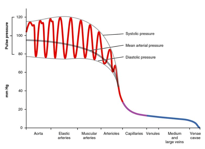
Figure i. The graph shows the components of claret pressure throughout the blood vessels, including systolic, diastolic, mean arterial, and pulse pressures.
Pulse Pressure
As shown in Figure 1, the difference between the systolic pressure and the diastolic pressure is the pulse pressure. For example, an individual with a systolic pressure level of 120 mm Hg and a diastolic pressure of fourscore mm Hg would have a pulse pressure level of 40 mmHg.
More often than not, a pulse pressure should exist at least 25 percentage of the systolic pressure. A pulse force per unit area below this level is described as low or narrow. This may occur, for example, in patients with a low stroke volume, which may be seen in congestive centre failure, stenosis of the aortic valve, or significant blood loss following trauma. In contrast, a high or wide pulse pressure level is common in salubrious people following strenuous do, when their resting pulse pressure of 30–40 mm Hg may increase temporarily to 100 mm Hg as stroke volume increases. A persistently high pulse pressure at or to a higher place 100 mm Hg may indicate excessive resistance in the arteries and can exist acquired past a variety of disorders. Chronic loftier resting pulse pressures can degrade the heart, brain, and kidneys, and warrant medical handling.
Mean Arterial Pressure
Mean arterial force per unit area (MAP) represents the "boilerplate" pressure of blood in the arteries, that is, the average strength driving blood into vessels that serve the tissues. Hateful is a statistical concept and is calculated by taking the sum of the values divided by the number of values. Although complicated to measure out directly and complicated to summate, MAP tin exist approximated by calculation the diastolic pressure level to one-third of the pulse force per unit area or systolic force per unit area minus the diastolic pressure level:
[latex]\text{MAP}=\text{diastolic BP}+\frac{(\text{systolic}-\text{diastolic BP})}{iii}[/latex]
In Figure 1, this value is approximately eighty + (120 − 80) / 3, or 93.33. Normally, the MAP falls within the range of 70–110 mm Hg. If the value falls below lx mm Hg for an extended time, blood pressure will not be high enough to ensure apportionment to and through the tissues, which results in ischemia, or bereft blood flow. A condition called hypoxia, inadequate oxygenation of tissues, commonly accompanies ischemia. The term hypoxemia refers to low levels of oxygen in systemic arterial blood. Neurons are especially sensitive to hypoxia and may die or be damaged if blood flow and oxygen supplies are not apace restored.
Pulse
Later on blood is ejected from the center, elastic fibers in the arteries help maintain a high-pressure gradient as they expand to accommodate the claret, so recoil. This expansion and recoiling event, known as the pulse, can be palpated manually or measured electronically. Although the effect diminishes over distance from the middle, elements of the systolic and diastolic components of the pulse are still evident down to the level of the arterioles.

Figure 2. The pulse is near readily measured at the radial artery, merely can be measured at any of the pulse points shown.
Because pulse indicates heart rate, it is measured clinically to provide clues to a patient'southward state of wellness. It is recorded equally beats per infinitesimal. Both the rate and the strength of the pulse are of import clinically. A high or irregular pulse rate tin can be acquired by physical activity or other temporary factors, merely it may also betoken a heart condition. The pulse forcefulness indicates the strength of ventricular contraction and cardiac output. If the pulse is strong, then systolic force per unit area is high. If it is weak, systolic pressure has fallen, and medical intervention may exist warranted.
Pulse can be palpated manually by placing the tips of the fingers across an artery that runs close to the body surface and pressing lightly. While this procedure is normally performed using the radial artery in the wrist or the common carotid artery in the cervix, whatever superficial avenue that can be palpated may be used. Common sites to find a pulse include temporal and facial arteries in the head, brachial arteries in the upper arm, femoral arteries in the thigh, popliteal arteries behind the knees, posterior tibial arteries virtually the medial tarsal regions, and dorsalis pedis arteries in the anxiety. A variety of commercial electronic devices are as well bachelor to measure pulse.
Measurement of Blood Force per unit area
Blood pressure is ane of the critical parameters measured on nigh every patient in every healthcare setting. The technique used today was developed more than 100 years ago by a pioneering Russian physician, Dr. Nikolai Korotkoff. Turbulent blood flow through the vessels can be heard as a soft ticking while measuring blood pressure; these sounds are known as Korotkoff sounds. The technique of measuring claret force per unit area requires the use of a sphygmomanometer (a blood pressure gage attached to a measuring device) and a stethoscope. The technique is as follows:
- The clinician wraps an inflatable gage tightly effectually the patient's arm at nigh the level of the heart.
- The clinician squeezes a prophylactic pump to inject air into the cuff, raising pressure around the artery and temporarilycutting off blood menses into the patient's arm.
- The clinician places the stethoscope on the patient'south antecubital region and, while gradually allowing air inside the gage to escape, listens for the Korotkoff sounds.
Although there are five recognized Korotkoff sounds, only ii are normally recorded. Initially, no sounds are heard since there is no blood catamenia through the vessels, only as air pressure drops, the cuff relaxes, and claret flow returns to the arm. As shown in Figure 3, the showtime sound heard through the stethoscope—the first Korotkoff audio—indicates systolic pressure. As more air is released from the cuff, blood is able to menstruation freely through the brachial artery and all sounds disappear. The point at which the concluding sound is heard is recorded every bit the patient's diastolic pressure.

Figure three. When pressure level in a sphygmomanometer cuff is released, a clinician can hear the Korotkoff sounds. In this graph, a blood pressure tracing is aligned to a measurement of systolic and diastolic pressures.
The bulk of hospitals and clinics have automated equipment for measuring claret pressure that work on the same principles. An even more recent innovation is a small instrument that wraps around a patient'southward wrist. The patient then holds the wrist over the heart while the device measures blood flow and records pressure (see Effigy ane).
Variables Affecting Blood Menstruation and Blood Pressure
Five variables influence blood flow and blood pressure:
- Cardiac output
- Compliance
- Volume of the blood
- Viscosity of the blood
- Blood vessel length and diameter
Retrieve that blood moves from college force per unit area to lower pressure. It is pumped from the centre into the arteries at high pressure. If y'all increase pressure level in the arteries (afterload), and cardiac function does not recoup, blood menstruum will actually decrease. In the venous arrangement, the reverse human relationship is truthful. Increased pressure in the veins does not decrease flow as it does in arteries, but actually increases flow. Since pressure in the veins is normally relatively depression, for blood to flow back into the heart, the pressure in the atria during atrial diastole must be even lower. It normally approaches zip, except when the atria contract.
Cardiac Output
Cardiac output is the measurement of blood flow from the heart through the ventricles, and is usually measured in liters per minute. Whatsoever gene that causes cardiac output to increase, past elevating heart rate or stroke volume or both, will elevate blood pressure and promote blood period. These factors include sympathetic stimulation, the catecholamines epinephrine and norepinephrine, thyroid hormones, and increased calcium ion levels. Conversely, any cistron that decreases cardiac output, by decreasing center charge per unit or stroke volume or both, volition decrease arterial pressure and blood flow. These factors include parasympathetic stimulation, elevated or decreased potassium ion levels, decreased calcium levels, anoxia, and acidosis.
Compliance
Compliance is the power of any compartment to aggrandize to adapt increased content. A metal pipe, for instance, is not compliant, whereas a balloon is. The greater the compliance of an artery, the more effectively it is able to expand to arrange surges in blood flow without increased resistance or blood pressure. Veins are more compliant than arteries and tin can expand to hold more than blood. When vascular illness causes stiffening of arteries, compliance is reduced and resistance to blood flow is increased. The outcome is more turbulence, higher pressure within the vessel, and reduced blood flow. This increases the work of the heart
A Mathematical Approach to Factors Affecting Blood Period
Jean Louis Marie Poiseuille was a French physician and physiologist who devised a mathematical equation describing blood menstruum and its relationship to known parameters. The same equation besides applies to engineering studies of the flow of fluids. Although understanding the math behind the relationships amongst the factors affecting blood flow is not necessary to understand claret flow, it can assist solidify an understanding of their relationships. Delight annotation that fifty-fifty if the equation looks intimidating, breaking it downwards into its components and following the relationships will make these relationships clearer, even if yous are weak in math. Focus on the iii critical variables: radius (r), vessel length (λ), and viscosity (η).
Poiseuille's equation:
[latex]\text{Blood flow}=\frac{\pi\Delta\text{Pr}^4}{8\eta\lambda}[/latex]
- π is the Greek alphabetic character pi, used to correspond the mathematical constant that is the ratio of a circumvolve's circumference to its diameter. Information technology may commonly be represented equally three.xiv, although the actual number extends to infinity.
- ΔP represents the difference in pressure.
- r4 is the radius (half of the diameter) of the vessel to the fourth ability.
- η is the Greek letter eta and represents the viscosity of the blood.
- λ is the Greek letter lambda and represents the length of a blood vessel.
One of several things this equation allows us to do is summate the resistance in the vascular system. Normally this value is extremely difficult to measure out, but it tin be calculated from this known relationship:
[latex]\text{Blood period}=\frac{\Delta\text{P}}{\text{Resistance}}[/latex]
If we rearrange this slightly,
[latex]\text{Resistance}=\frac{\Delta\text{P}}{\text{Blood flow}}[/latex]
Then by substituting Pouseille'due south equation for blood menstruum:
[latex]\text{Resistance}=\frac{8\eta\lambda}{\pi\text{r}^4}[/latex]
By examining this equation, you can see that in that location are only three variables: viscosity, vessel length, and radius, since 8 and π are both constants. The important affair to remember is this: Two of these variables, viscosity and vessel length, will alter slowly in the body. Just ane of these factors, the radius, tin can be changed speedily by vasoconstriction and vasodilation, thus dramatically impacting resistance and flow. Further, pocket-size changes in the radius will greatly impact flow, since information technology is raised to the fourth power in the equation.
We accept briefly considered how cardiac output and claret volume impact blood flow and pressure level; the next step is to see how the other variables (contraction, vessel length, and viscosity) articulate with Pouseille's equation and what they tin can teach usa virtually the impact on blood flow.
Blood Volume
The relationship betwixt claret volume, claret force per unit area, and claret period is intuitively obvious. Water may merely trickle along a creek bed in a dry season, but rush quickly and under smashing pressure afterwards a heavy rain. Similarly, equally blood book decreases, pressure and flow subtract. As blood volume increases, pressure and menstruum increase.
Under normal circumstances, blood volume varies little. Low blood volume, called hypovolemia, may exist caused by bleeding, dehydration, vomiting, astringent burns, or some medications used to treat hypertension. It is important to recognize that other regulatory mechanisms in the trunk are so effective at maintaining blood pressure that an individual may be asymptomatic until 10–20 per centum of the claret volume has been lost. Handling typically includes intravenous fluid replacement.
Hypervolemia, excessive fluid volume, may be caused past retention of h2o and sodium, as seen in patients with center failure, liver cirrhosis, some forms of kidney disease, hyperaldosteronism, and some glucocorticoid steroid treatments. Restoring homeostasis in these patients depends upon reversing the condition that triggered the hypervolemia.
Blood Viscosity
Viscosity is the thickness of fluids that affects their ability to menses. Make clean water, for example, is less sticky than mud. The viscosity of blood is directly proportional to resistance and inversely proportional to flow; therefore, any condition that causes viscosity to increase will besides increase resistance and subtract menses. For example, imagine sipping milk, then a milkshake, through the same size straw. Y'all experience more resistance and therefore less flow from the milkshake. Conversely, any condition that causes viscosity to decrease (such every bit when the milkshake melts) will decrease resistance and increase flow.
Unremarkably the viscosity of blood does not change over brusk periods of time. The two primary determinants of blood viscosity are the formed elements and plasma proteins. Since the vast majority of formed elements are erythrocytes, any condition affecting erythropoiesis, such as polycythemia or anemia, can alter viscosity. Since about plasma proteins are produced by the liver, any status affecting liver part can also change the viscosity slightly and therefore decrease blood flow. Liver abnormalities include hepatitis, cirrhosis, alcohol damage, and drug toxicities. While leukocytes and platelets are normally a small component of the formed elements, there are some rare atmospheric condition in which severe overproduction can impact viscosity too.
Vessel Length and Diameter
The length of a vessel is direct proportional to its resistance: the longer the vessel, the greater the resistance and the lower the catamenia. As with blood book, this makes intuitive sense, since the increased surface expanse of the vessel volition impede the flow of blood. Likewise, if the vessel is shortened, the resistance will subtract and catamenia will increase.
The length of our blood vessels increases throughout babyhood as we grow, of course, but is unchanging in adults nether normal physiological circumstances. Farther, the distribution of vessels is non the same in all tissues. Adipose tissue does not have an all-encompassing vascular supply. One pound of adipose tissue contains approximately 200 miles of vessels, whereas skeletal muscle contains more than twice that. Overall, vessels subtract in length only during loss of mass or amputation. An private weighing 150 pounds has approximately 60,000 miles of vessels in the body. Gaining near ten pounds adds from 2000 to 4000 miles of vessels, depending upon the nature of the gained tissue. One of the cracking benefits of weight reduction is the reduced stress to the heart, which does not have to overcome the resistance of as many miles of vessels.
In contrast to length, the bore of blood vessels changes throughout the body, according to the type of vessel, as we discussed earlier. The diameter of any given vessel may as well alter oft throughout the twenty-four hours in response to neural and chemic signals that trigger vasodilation and vasoconstriction. The vascular tone of the vessel is the contractile state of the smooth musculus and the primary determinant of diameter, and thus of resistance and flow. The consequence of vessel diameter on resistance is changed: Given the same volume of claret, an increased diameter means there is less blood contacting the vessel wall, thus lower friction and lower resistance, subsequently increasing menstruum. A decreased diameter means more of the claret contacts the vessel wall, and resistance increases, subsequently decreasing flow.
The influence of lumen diameter on resistance is dramatic: A slight increase or decrease in diameter causes a huge subtract or increment in resistance. This is because resistance is inversely proportional to the radius of the blood vessel (half of the vessel's bore) raised to the 4th ability (R = 1/r4). This means, for example, that if an artery or arteriole constricts to one-half of its original radius, the resistance to flow will increase sixteen times. And if an artery or arteriole dilates to twice its initial radius, then resistance in the vessel volition subtract to 1/sixteen of its original value and flow will increase sixteen times.
The Roles of Vessel Diameter and Total Expanse in Blood Flow and Claret Pressure level
Recall that we classified arterioles every bit resistance vessels, because given their small lumen, they dramatically boring the period of claret from arteries. In fact, arterioles are the site of greatest resistance in the entire vascular network. This may seem surprising, given that capillaries take a smaller size. How can this phenomenon be explained?
Figure 4 compares vessel diameter, full cross-sectional area, boilerplate blood force per unit area, and blood velocity through the systemic vessels. Notice in parts (a) and (b) that the full cross-sectional area of the torso's capillary beds is far greater than any other type of vessel. Although the diameter of an individual capillary is significantly smaller than the diameter of an arteriole, there are vastly more capillaries in the body than there are other types of blood vessels. Part (c) shows that blood force per unit area drops unevenly as claret travels from arteries to arterioles, capillaries, venules, and veins, and encounters greater resistance. However, the site of the most precipitous drop, and the site of greatest resistance, is the arterioles. This explains why vasodilation and vasoconstriction of arterioles play more meaning roles in regulating claret pressure than do the vasodilation and vasoconstriction of other vessels.

Figure four. The relationships among blood vessels that can be compared include (a) vessel bore, (b) total cross-exclusive area, (c) boilerplate blood pressure, and (d) velocity of blood flow.
Role (d) shows that the velocity (speed) of blood period decreases dramatically as the blood moves from arteries to arterioles to capillaries. This slow flow rate allows more time for exchange processes to occur. As blood flows through the veins, the rate of velocity increases, equally blood is returned to the heart.
Disorders of the Cardiovascular System: Arteriosclerosis
Compliance allows an artery to aggrandize when blood is pumped through it from the heart, and and then to recoil after the surge has passed. This helps promote blood menstruation. In arteriosclerosis, compliance is reduced, and pressure and resistance within the vessel increase. This is a leading cause of hypertension and coronary center disease, every bit it causes the center to work harder to generate a pressure dandy plenty to overcome the resistance.
Arteriosclerosis begins with injury to the endothelium of an artery, which may exist caused by irritation from high blood glucose, infection, tobacco employ, excessive blood lipids, and other factors. Artery walls that are constantly stressed by claret flowing at high pressure are also more than likely to be injured—which means that hypertension tin promote arteriosclerosis, as well as event from it.
Think that tissue injury causes inflammation. As inflammation spreads into the artery wall, it weakens and scars it, leaving it potent (sclerotic). As a result, compliance is reduced. Moreover, circulating triglycerides and cholesterol can seep betwixt the damaged lining cells and become trapped within the artery wall, where they are frequently joined by leukocytes, calcium, and cellular debris. Eventually, this buildup, called plaque, can narrow arteries plenty to impair blood menstruum. The term for this condition, atherosclerosis (athero- = "porridge") describes the mealy deposits.
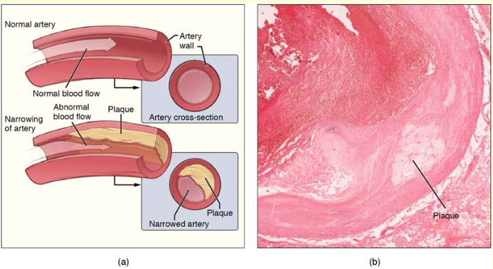
Effigy 5. Atherosclerosis. (a) Atherosclerosis can event from plaques formed past the buildup of fatty, calcified deposits in an avenue. (b) Plaques can also take other forms, as shown in this micrograph of a coronary artery that has a buildup of connective tissue within the artery wall. LM × 40. (Micrograph provided past the Regents of University of Michigan Medical School © 2012)
Sometimes a plaque can rupture, causing microscopic tears in the artery wall that allow blood to leak into the tissue on the other side. When this happens, platelets rush to the site to clot the blood. This jell tin farther obstruct the artery and—if information technology occurs in a coronary or cerebral avenue—cause a sudden heart attack or stroke. Alternatively, plaque tin can intermission off and travel through the bloodstream as an embolus until it blocks a more distant, smaller artery.
Even without total blockage, vessel narrowing leads to ischemia—reduced claret menstruation—to the tissue region "downstream" of the narrowed vessel. Ischemia in turn leads to hypoxia—decreased supply of oxygen to the tissues. Hypoxia involving cardiac musculus or brain tissue tin lead to prison cell death and severe impairment of encephalon or middle function.
A major risk gene for both arteriosclerosis and atherosclerosis is advanced age, as the conditions tend to progress over fourth dimension. Arteriosclerosis is normally divers equally the more generalized loss of compliance, "hardening of the arteries," whereas atherosclerosis is a more specific term for the build-upward of plaque in the walls of the vessel and is a specific type of arteriosclerosis. There is as well a distinct genetic component, and pre-existing hypertension and/or diabetes also greatly increase the adventure. Nevertheless, obesity, poor nutrition, lack of physical activity, and tobacco use all are major risk factors.
Handling includes lifestyle changes, such as weight loss, smoking cessation, regular exercise, and adoption of a diet low in sodium and saturated fats. Medications to reduce cholesterol and claret pressure level may be prescribed. For blocked coronary arteries, surgery is warranted. In angioplasty, a catheter is inserted into the vessel at the point of narrowing, and a second catheter with a balloon-similar tip is inflated to widen the opening. To prevent subsequent collapse of the vessel, a small mesh tube called a stent is ofttimes inserted. In an endarterectomy, plaque is surgically removed from the walls of a vessel. This operation is typically performed on the carotid arteries of the neck, which are a prime source of oxygenated blood for the brain. In a coronary bypass process, a non-vital superficial vessel from another part of the body (oftentimes the dandy saphenous vein) or a constructed vessel is inserted to create a path effectually the blocked area of a coronary artery.
Venous System
The pumping action of the heart propels the blood into the arteries, from an surface area of higher pressure toward an area of lower pressure level. If blood is to catamenia from the veins back into the heart, the force per unit area in the veins must be greater than the pressure in the atria of the center. Two factors assistance maintain this pressure gradient between the veins and the centre. Starting time, the pressure in the atria during diastole is very depression, oft approaching goose egg when the atria are relaxed (atrial diastole). Second, two physiologic "pumps" increment pressure in the venous system. The utilize of the term "pump" implies a physical device that speeds flow. These physiological pumps are less obvious.
Skeletal Muscle Pump
In many body regions, the pressure inside the veins tin can exist increased by the contraction of the surrounding skeletal muscle. This mechanism, known as the skeletal muscle pump (Figure 6), helps the lower-pressure veins counteract the forcefulness of gravity, increasing pressure to move blood dorsum to the eye. As leg muscles contract, for case during walking or running, they exert pressure on nearby veins with their numerous ane-way valves. This increased pressure causes blood to flow upwardly, opening valves superior to the contracting muscles so blood flows through. Simultaneously, valves inferior to the contracting muscles shut; thus, blood should not seep back downward toward the anxiety. Armed forces recruits are trained to flex their legs slightly while continuing at attending for prolonged periods. Failure to do and then may allow blood to puddle in the lower limbs rather than returning to the heart. Consequently, the brain volition not receive enough oxygenated claret, and the individual may lose consciousness.

Figure 6. The contraction of skeletal muscles surrounding a vein compresses the blood and increases the pressure in that area. This action forces blood closer to the center where venous pressure is lower. Note the importance of the 1-fashion valves to assure that claret flows only in the proper management.
Respiratory Pump
The respiratory pump aids blood flow through the veins of the thorax and abdomen. During inhalation, the volume of the thorax increases, largely through the contraction of the diaphragm, which moves downward and compresses the abdominal cavity. The elevation of the chest caused by the contraction of the external intercostal muscles also contributes to the increased book of the thorax. The volume increase causes air force per unit area within the thorax to decrease, allowing us to inhale. Additionally, every bit air force per unit area within the thorax drops, claret pressure in the thoracic veins also decreases, falling beneath the pressure in the abdominal veins. This causes blood to catamenia forth its pressure slope from veins outside the thorax, where pressure is college, into the thoracic region, where pressure is now lower. This in turn promotes the return of blood from the thoracic veins to the atria. During exhalation, when air pressure level increases within the thoracic cavity, pressure level in the thoracic veins increases, speeding blood menstruum into the heart while valves in the veins foreclose claret from flowing astern from the thoracic and abdominal veins.
Pressure Relationships in the Venous System
Although vessel diameter increases from the smaller venules to the larger veins and eventually to the venae cavae (singular = vena cava), the total cross-sectional area actually decreases. The individual veins are larger in diameter than the venules, simply their full number is much lower, so their full cantankerous-sectional area is also lower.
Also notice that, every bit blood moves from venules to veins, the average blood pressure level drops, but the blood velocity actually increases. This pressure gradient drives blood back toward the heart. Over again, the presence of 1-way valves and the skeletal muscle and respiratory pumps contribute to this increased flow. Since approximately 64 percent of the total blood volume resides in systemic veins, whatever action that increases the catamenia of blood through the veins volition increase venous render to the eye. Maintaining vascular tone within the veins prevents the veins from merely distending, dampening the flow of blood, and as yous will see, vasoconstriction actually enhances the flow.
The Function of Venoconstriction in Resistance, Blood Pressure level, and Flow
As previously discussed, vasoconstriction of an artery or arteriole decreases the radius, increasing resistance and pressure, but decreasing flow. Venoconstriction, on the other hand, has a very different issue. The walls of veins are thin but irregular; thus, when the polish muscle in those walls constricts, the lumen becomes more rounded. The more rounded the lumen, the less surface expanse the claret encounters, and the less resistance the vessel offers. Vasoconstriction increases force per unit area inside a vein equally information technology does in an avenue, but in veins, the increased pressure increases flow. Recall that the pressure in the atria, into which the venous blood volition catamenia, is very low, approaching nil for at least part of the relaxation stage of the cardiac bike. Thus, venoconstriction increases the return of claret to the eye. Another way of stating this is that venoconstriction increases the preload or stretch of the cardiac musculus and increases contraction.
Affiliate Review
Blood menstruation is the motion of blood through a vessel, tissue, or organ. The slowing or blocking of blood menstruum is chosen resistance. Blood force per unit area is the forcefulness that claret exerts upon the walls of the blood vessels or chambers of the heart. The components of blood pressure include systolic pressure level, which results from ventricular wrinkle, and diastolic pressure, which results from ventricular relaxation. Pulse force per unit area is the difference between systolic and diastolic measures, and hateful arterial pressure is the "average" pressure of blood in the arterial system, driving blood into the tissues. Pulse, the expansion and recoiling of an artery, reflects the heartbeat. The variables affecting blood flow and blood force per unit area in the systemic circulation are cardiac output, compliance, claret volume, blood viscosity, and the length and diameter of the blood vessels. In the arterial system, vasodilation and vasoconstriction of the arterioles is a meaning gene in systemic blood pressure: Slight vasodilation profoundly decreases resistance and increases flow, whereas slight vasoconstriction greatly increases resistance and decreases menstruum. In the arterial arrangement, as resistance increases, blood pressure increases and flow decreases. In the venous system, constriction increases claret pressure every bit information technology does in arteries; the increasing pressure level helps to return claret to the heart. In addition, constriction causes the vessel lumen to get more rounded, decreasing resistance and increasing claret flow. Venoconstriction, while less important than arterial vasoconstriction, works with the skeletal muscle pump, the respiratory pump, and their valves to promote venous return to the eye.
Self Cheque
Answer the question(s) below to see how well yous understand the topics covered in the previous section.
Disquisitional Thinking Questions
- Y'all take a patient'southward blood pressure, it is 130/ 85.
Calculate the patient'southward pulse pressure and mean arterial pressure. Decide whether each pressure is low, normal, or high. - An obese patient comes to the clinic lament of bloated anxiety and ankles, fatigue, shortness of breath, and oft feeling "spaced out." She is a cashier in a grocery store, a job that requires her to stand all twenty-four hours. Outside of work, she engages in no physical activity. She confesses that, considering of her weight, she finds even walking uncomfortable. Explicate how the skeletal muscle pump might play a office in this patient's signs and symptoms.

Source: https://courses.lumenlearning.com/suny-ap2/chapter/blood-flow-blood-pressure-and-resistance-no-content/
Posted by: gasparhossing.blogspot.com
0 Response to "Which Of The Following Vessels Changes Diameter Most Readily To Local Conditions Or Sns Stimulation?"
Post a Comment